A torn ACL is one of the most common injuries that can occur on the body. Whether you’ve torn your ACL by playing sports or through another form of physical activity, the pain can be excruciating. To determine whether you have a torn ACL or a different type of knee injury, your doctor might recommend an MRI exam.
What is a Torn ACL?
A torn ACL is a tear of the anterior cruciate ligament (ACL) which is one of the major ligaments in the knee. The ACL is located inside your knee joint where the femur, tibia, and kneecap join together. A torn ACL commonly occurs during physical activity that involves a sudden change of direction or a quick stop. Sports such as football, soccer, downhill skiing, and basketball can all see common torn ACL occurrences.
ACL injuries include mild sprains, partial and complete tears. A mild sprain means the ligament is still working, but it’s been stretched out of place. A partial tear is when the ACL has been stretched so far, it’s now fully loose. A complete ACL tear is when the ligament is torn into two pieces and the knee joint is now unstable.
Symptoms for a torn ACL can start with an audible popping noise in the knee. Afterward, your knee may begin to swell, you can lose your range of motion, and your knee may become too painful to bear your full weight.
It’s important to seek immediate care if your knee is showing signs of a torn ACL. An accurate diagnosis can help doctors determine the severity of the injury and provide you with the proper treatment.
Torn ACL MRI Procedure
A knee MRI uses magnetic fields and radio waves to create images of the inside of your knee. To prepare for the torn ACL MRI procedure, let your doctor know if you have any metal inside your body that could impact the MRI exam. Metal jewelry or other items on your body may need to be removed so as not to disrupt the process. Unless otherwise advised by a doctor, you can go about your normal routine of eating and drinking before taking an exam.
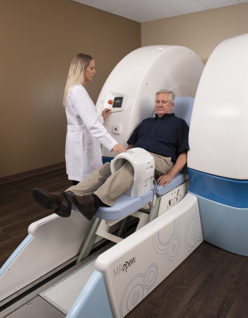
With a traditional closed MRI machine, you rest completely flat on a cushioned table that moves into the scanner so your technologist can capture images of your knee. It’s important to be as still as possible so the highest quality of images are processed. An open MRI helps eliminate the claustrophobic feel that some people struggle with during a closed MRI. An open MRI also provides more comfort to larger patients. During an open MRI, you will either sit or stand determining how the technologist wants to view your injury.
Once your MRI exam is complete, the MRI software transfers the images to a HIPAA compliant computer. A radiologist reviews the images on a high-resolution monitor to assess the joint and surrounding tissue to determine whether or not you have a torn ACL.
At American Health Imaging, we have the latest high-quality MRI equipment and expertly trained radiologists to help you quickly determine if you have a torn ACL. If you are experiencing pain in your knee, it’s important to get a diagnosis as soon as possible so you can take the next best steps toward recovery.
Contact us today or talk to your doctor about scheduling your MRI with us so we can help you determine the best course of action for your knee injury.